


Introduction
Heart rate change is frequently seen in epileptic seizures, for example, ictal tachycardia and ictal bradycardia. Ictal tachycardia is defined as the occurrence of sinus tachycardia around the onset of ictal discharges.1 The prevalence of ictal tachycardia is very common(80–100%).2–5 It occurs more frequently in seizures with temporal lobe origin,6,7 especially with mesial temporal lobe onset.6,8 Ictal bradycardia is less frequent than ictal tachycardia; the prevalence has been calculated as ranging from 2.1% to 25.5%.4,9 It is most prevalent in seizures of the temporal lobe origin,9,10 and has a stronger association with bilateral hemispheric seizures.11
The heart rate (HR) is one of the most easy way to detect autonomic activity. The autonomic system (parasympathetic and sympathetic systems) has a major role to maintain homeostasis and regulate visceral functions such as HR.12 Epileptic seizures can cause changes in the autonomic functions affecting the sympathetic, parasympathetic, and enteric nervous systems.12 Epileptic seizure affects autonomic functions chemically or electrically.13 The anterior cingulate, insular, posterior orbito-frontal, and the pre-frontal cortices have important roles in regulating the autonomic nervous system at the cortical level; the amygdala, thalamus, and hypothalamus influencing autonomic nervous system at the subcortical level.1,14–16 In patients with epilepsy, the ictal discharges are propagated to these structures and the sympathetic or parasympathetic outflows are increased and this results in autonomic changes, such as a tachycardia.12 Tachycardia is earlier in right temporal lobe epilepsy (TLE) than left TLE because of right cerebral hemisphere is dominant in the sympathetic network.17
It is important extinguish from TLE to frontal lobe epilepsy (FLE). There are many studies comparing TLE and FLE. But those studies were focused on clinical semiology, such as the aura, automatisms, vocalization, and secondarily generalization or post-ictal symptoms. There were some studies about the HR change between TLE and extra temporal lobe epilepsy (XTLE),6,18,19 Adjei et al. investigated during subclinical electrographic seizures,18 but there has been no study about the difference of autonomic features, such as amount of change of HR, maximal HR, duration on HR change and so on, between TLE and FLE during clinical seizure - to the best of our knowledge. So, we compared the autonomic function or heart rate changes during seizures between FLE and TLE.
Methods
Patients and seizures
We retrospectively reviewed the medical records of the Samsung Medical Center epilepsy monitoring unit (Seoul, South Korea) of those patients who had underwent long-term video EEG monitoring between January 2014 and April 2015. The inclusion criteria for this study were: (1) tachycardia, the HR exceed 100 bpm or increased 20% from baseline HR, (2) partial seizures without secondarily generalization, (3) scalp EEG seizures arising from the temporal or frontal regions, (4) identifiable QRS complexes on ictal ECG, and (5) more than 180 seconds of intervals with previous seizure.
The exclusion criteria for this study were: (1) any other diseases influencing autonomic system, (2) neurologic deficit, (3) ECG abnormality during the interictal period, (4) the seizures were emerged before 180 sec from previous seizure.
EEG and ECG recording
A scalp EEG was performed using the 10–20 system with additional frontotempral electrodes, simultaneous with ECG, right shoulder, left shoulder, A1, and A2 electrodes in an epilepsy monitoring unit (EMU). The data were recorded by the NicoletOne LTM system (Natus Medical Incorporated, Pleasanton, CA, USA) with a sampling rate of 512Hz. The EEG seizure onset was determined by three board certified neurologists.
Preictal-ictal-postictal HR changes
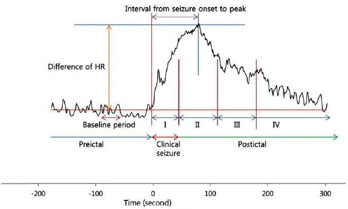
The HR was calculated form ECG R-R intervals or S-S intervals. The R-R intervals or S-S intervals were extracted by Microsoft Excel (MS Office 2010, Microsoft, USA). We obtained the ECG data during 180 seconds before the seizure onset, ictal period, and 180 seconds after the end of the seizure. The methods of this study can be seen in Figure 1. The “baseline HR” was determined by the mean HR from 120 seconds before a seizure onset to 90 seconds before a seizure onset. The “maximum HR” was determined by the biggest HR, after correction for the artifacts. The “seizure onset to peak” was defined as the time between the seizure onset and the time at maximum HR. The “HR change” was defined as the difference between the maximum HR and baseline HR. The “return to baseline” was defined as the time between the time of maximum HR and the time that the HR became smaller than the baseline HR after the maximum HR. The “return to baseline” were categorized as group 1; the return to baseline during ictal period as group 2; within 60 seconds after the ictal periods as group 3; between 60 seconds and 120 seconds after the ictal periods as group 4; and more than 120 seconds after ictal periods.
Statistical analysis
There was a comparison between FLE and TLE concerning the seizure duration, baseline HR, maximum HR, HR change, seizure onset to peak, and return to baseline. The statistical assessments were performed with the generalized estimating equation using SAS version 9.4 (SAS institute Inc, Cary, NC, USA)
Results
From January, 2014 to April, 2015, there were 391 patients admitted at EMU. 51 patients had tachycardia. We selected 10 TLE, 10 FLE with matching sex. A total of 20 patients were enrolled in this study; 10 patients were men and 10 were women, aged 15 to 66 years. There were 10 patients who were FLE, with 10 as TLE. In total, 198 seizures were analyzed, 83 were TLE and 115 were FLE; with 87 seizures being right oriented, and 18 were left, 93 were bilateral. Table 1 shows the patients, seizure type, and onset. Figure 2 shows some typical examples of heart rate around the seizures in patients with TLE and FLE. The change of HR is prominent and prolonged in TLE than FLE. Table 2 shows the difference between FLE and TLE. The mean duration of seizures in TLE was 93.80 ± 54.92 seconds, whereas in FLE it was 39.0 ± 21.4 seconds, with a difference of 54.8 seconds (p < 0.001). The baseline HR was similar for the two groups: 74.9 ± 17.2 and 70.70 ± 11.5 bpm. The time to peak HR was 135.1 ± 19.1 seconds in the TLE group, whereas it was 119.3 ± 19.7 seconds in the FLE group, with a difference of 15.8 seconds (p = 0.027). The HR change was 60.0 ± 26.3 bpm in the TLE group, whereas it was 48.7 ± 18.5 bpm in the FLE group, with a difference of 11.3 seconds (p < 0.001). The mean duration of the seizure onset to peak was 47.8 ± 45.4 seconds in the TLE group, whereas it was 22.8 ± 26.2 seconds in the FLE group, with a difference of 25.0 (p < 0.001). Figure 3 shows the difference of the return to baseline between TLE and FLE groups. The seizures that returned to the baseline during the ictal period were 1 in the TLE group (1.2%) versus 9 in the FLE (7.83%) group. Before 60 seconds there were 19 in the TLE group (22.89%), whereas there were 62 in the FLE group (53.91%). Between 60 seconds and 120 seconds, there were 5 in the TLE group (6.02%), with 15 in the FLE group (13.04%). Over 120 seconds, there were 58 in the TLE (69.88%) group, whereas there were 29 in the FLE group (25.22%). Thus, in brief summary, in the TLE group, many seizures (69.88%) were returned to the baseline HR longer than 120 seconds, whereas in the FLE group, most (61.74%) were before 60 seconds. The p-value was 0.001.
Discussion
The results of this study revealed that ictal tachycardia was observed in both the FLE and TLE group, but the HR change was different between TLE group and FLE group. We can suggest the reasons why TLE group has more prominent and prolonged than FLE group, the limbic system, which includes the amygdala and the hippocampus, and the insular cortex, are keys to the autonomic regulation in epilepsy. Tachycardia is more frequent in the TLE than other seizures, because of the involvement of the amygdala, hippocampus and right insular cortex.7 Many studies suggest that the propagation of epileptic discharges to the right insular cortex is a key part of the sympathetic-parasympathetic changes that influences the heart rate.20 Ictal tachycardia is more prominent in temporal lobe epilepsy (TLE),21 as compared with XTLE,9,22 which may suggest that the involvement of insular cortex is a key part of ictal tachycardia.1 The insular has a role in regulating autonomic nervous system.23 In TLE, compared with XTLE, the duration of the tachycardia is longer. This may be because epileptic discharges can propagate toward the insular cortex more easily and longer in TLE as compared with XTLE.1 There was tachycardia in FLE, the reason of tachycardia of FLE is due to orbitofrontal cortex.
Autonomic suprabulbar influences are mediated by several output pathways from orbitofrontal cortex to the thalamus, temporal lobe, amygdala, brain stem and so on.6 Stimulation of frontocortical-brain stem pathways influences sympathetic tone increase and may produce cardiac arrhythmias.24 Thus, the difference of HR between FLE and TLE was because of the involvement of the limbic system or orbitofrontal system. Compare TLE and FLE, the limbic system and insular cortex has more profound effect to the autonomic system than orbitofrontal cortex.
Tachycardia is one of the most important risk factor of sudden unexpected death in epilepsy (SUDEP).25 Tachycardia may be considered one of the key event in tachyarrhythmia and sympathetic over-activity. Tachyarrhythmia alter cardiac repolarization, and that make critical arrhythmia, and finally makes cardiac arrest.26 In TLE, more patient has higher risk of SUDEP.27 In our study, there are more prolonged and prominent HR change in TLE, and there are more sympathetic over-activity, and have more chance of cardiac arrhythmia, and so the patients might have higher risk of SUDEP.
The limitation of present study is the small number of patients, as well as the small numbers of FLE from the left hemisphere. We collected the data from an epilepsy monitoring unit, which estimated the benefit and possibility of epilepsy surgery. The left FLE has less chance to be operated on because of the presence of the language center in the left frontal lobe. Thus, very few left FLE patients and seizures are included in this study. Hence, more studies are needed to clarify the difference in HR pattern between FLE and TLE.
Other studies have been about ictal tachycardia, which focused on the start of the HR change and ictal onset. So, for example, they published which epileptic syndrome is earlier than another.17 Many studies analyzed the onset of HR change, seizure onset, and the correlation between them, but in this present study, there were many motion artifacts, thus we could not find the point to change the HR.
In TLE, the HR change is much prominent and prolonged than FLE, because of ictal discharge propagation to the limbic system and insular regions. Therefore, this difference can be an important point to differentiate between FLE and TLE.