Introduction
Transient amnesia is a commonly encountered clinical manifestation with diverse causes. The most common cause of transient amnesia is transient global amnesia (TGA), which is characterized by the abrupt onset of anterograde amnesia without disturbances in other cognitive functions. TGA usually lasts for a few hours and is accompanied by typical behavioral changes.1,2 Transient epileptic amnesia (TEA) is a sub-type of mesial temporal lobe epilepsy characterized by recurrent episodes of transient memory disturbances. TEA closely resembles TGA because transient amnesia is sometimes the sole manifestation of the seizures. Although TEA is distinguished from TGA by a relatively brief duration (less than an hour), high frequency of episodes, common occurrence upon waking, patients with TEA generally cannot remember the amnestic episodes.2–5
Here we report the cases of two patients with TEA who presented with amnesia with preserved consciousness. They were amnesic during the episodes, but remembered the events after the episodes. These episodes were caused by non-dominant temporal lobe epilepsy. We will then discuss the mechanisms underlying our observations.
Case
Case 1
A 77-year-old right-handed man was admitted to our department with recurrent episodes of transient amnesia. The patient had no other notable past medical or psychiatric disease. Two days before admission, the patient experienced an episode of transient amnesia that began abruptly during breakfast. He could not remember his job and kept asking why he had to go to work despite his wife’s repeated explanations. The symptoms lasted for about 10 minutes and then disappeared. Similar symptoms were observed again on the next day. The patient did not remember his nephew’s wedding, which had taken place 4 days prior. He continued to ask when the nephew was married and why he was married so early. The patient’s memories gradually returned over a period of 20 minutes. After these episodes, however, the patient remembered what happened during those events. During the above episodes, the patient’s family did not observe any other cognitive dysfunction other than amnesia. The patient was alert, fluent, and not disoriented to place or person.
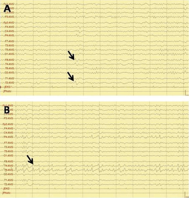
Routine physical and neurological examination revealed no abnormalities. The patient had a score of 29 on the Korean version of the mini-mental examination (K-MMSE) (2 out of 3 on memory recall test, could recall the other when cued). Magnetic resonance imaging (MRI) and MR angiography revealed no abnormalities in the hippocampi or other structures related to memory. On electroencephalographic (EEG), epileptiform discharges were observed in the right temporal leads, including F8 and T2 (Fig. 1A). Brain single-photon emission computed tomography revealed no significant increase or decrease of perfusion in the temporal lobes. Routine blood tests, including blood cell counts, routine chemistry tests, and tests to determine the levels of ammonia and creatine phosphokinase revealed no significant abnormalities. The patient was treated with 15 mg/kg of carbamazepine controlled-release (CR), followed by maintenance dose of 200 mg twice per day. He has been free of memory disturbances for 3 months.
Case 2
A 63-year-old right-handed woman was admitted for evaluation of recurrent and brief memory disturbances. Her husband noticed that the patient suffered from retrograde amnesia upon waking. Specifically, the patient repeatedly asked about what she did last night. The amnesia lasted for about thirty minutes. During these attacks, cognitive functions other than memory seemed to remain intact. One month later, when the patient exited the subway train, she lost her way for about 10 minutes. The patient remembered these amnesic episodes and what she did during those events.
Routine physical and neurological examination revealed no abnormalities. The patient had a score of 24 on the K-MMSE (1 out of 5 on the attenuation and concentration test and 1 out of 3 on the memory recall test, could recall the others when cued). The patient did not show significant declines in cognitive functions and activities of daily living observed by her family. Brain MRI, MR angiogram, and EEG revealed no abnormalities. Routine blood tests also revealed no abnormalities. The patient was treated with 300 mg of valproic acid twice per day. She had not been taking any medicine about cognitive dysfunction before and after those tests. Her symptoms did not recur for 3 years.
However, she developed transient repeated amnesia after the symptom-free interval. Diffusion-weighted MRI and blood tests revealed no abnormalities, but intermittent rhythmic high-voltage sharp waves were observed in the right temporal area (Fig. 1B). After the addition of 100 mg zonisamide to her medication, the patient has been symptom-free for 1 month.
Discussion
TEA is a rare but distinct temporal lobe epilepsy, and is characterized by recurrent ictal transient memory disturbances. These disturbances typically occur upon waking. TEA is often accompanied by other temporal seizures, such as olfactory hallucinations and oral automatisms. It responds favorably to anti-epileptic medication.3–5 Until now, the reported EEG findings have mostly been interictal. These findings include unilateral or bilateral temporal sharp waves, nonspecific focal slow waves, or are normal.6 EEG abnormalities areusually observed in the dominant side or in bilateral areas.
Our patients had many features compatible with the diagnostic criteria proposed by Zeman and Bulter.4 Specifically, they had histories of recurrent episodes of transient amnesia lasting for less than an hour and responded well to anti-epileptic medication. In addition, their cognitive abilities, such as perception and language, were intact during the episodes.4
TEA is easily mistaken for TGA or psychogenic amnesia. However, episodes of TGA tend to last several hours and have lower recurrence rates, while episodic memory of psychogenic amnesia remains intact and the acquisition for new memories is preserved.1,2,7 Thus, both of these conditions differ from those seen in our patients.
Our patients had atypical clinical and EEG findings. Specifically, they remembered the amnesic episodes. Few patients with TEA can remember their attacks in detail.6 Interestingly, these patients displayed repetitive questioning during the TEA attacks. Another interesting observation was that abnormal discharges on EEG were detected on the temporal lobe of the right hemisphere (speech/non-dominant side), but not on the dominant side or bilaterally.
It has been reported that the lateralization of temporal lobe seizures may be the predominant feature determining whether consciousness is preserved ictally. The dominant hemisphere is primarily responsible for the conscious state.8–10 Consciousness impairments are more often observed in patients with dominant temporal seizures than in patients with seizures in non-dominant areas.11 In addition, several reports indicate that consciousness is preserved at seizure onset as long as synchronization remains confined to the non-dominant temporal lobe. Loss of consciousness is thought to be directly related to excessive synchronization of neuronal activity in extra-temporal lobe structures, including the thalamus and parietal cortices.12–14 We thus hypothesize our patients were able to recollect the amnestic episode itself after the episode because ictal rhythms originating from the non-dominant temporal lobe did not propagate to extra-temporal structures, such as the thalamus and associated cortices, which are involved in loss of consciousness during the attack.
In summary, we report two cases of TEA with atypical features, including recollection of the amnestic episode and abnormal discharges in the non-dominant temporal lobe. We propose that confinement of epileptic discharge to the non-dominant temporal lobe can lead to partial preservation of consciousness during seizure attacks that produce transient amnesia.


