Posterior Reversible Encephalopathy Syndrome Due to Hyponatremia
Article information
Abstract
Posterior reversible encephalopathy syndrome (PRES) is characterized by variable associations of seizure activity, consciousness impairment, headaches, visual abnormalities, nausea/vomiting, and focal neurological signs. The PRES may occur in diverse situations. The findings on neuroimaging in PRES are often symmetric and predominate edema in the white matter of the brain areas perfused by the posterior brain circulation, which is reversible when the underlying cause is treated. We report the case of PRES in normotensive patient with hyponatremia.
Introduction
Posterior reversible encephalopathy syndrome (PRES) was initially described in 1996 by Hinchey and co-workers, as reversible posterior leukoencephalopathy syndrome.1 It is characterized by variable associations of seizure activity, consciousness impairment, headaches, visual abnormalities, nausea/vomiting, and focal neurological signs.2 The term PRES describes a potentially reversible imaging appearance and may occur in diverse situations. While most cases are due to systemic hypertension (HTN), other conditions and entities have been identified as etiologic or risk factors in the absence of HTN, such as immunosuppressant drugs use, nephrotic state, sepsis, and systemic lupus erythematosus (SLE).2–4 The findings on neuroimaging in PRES are often symmetric and predominate edema in the white matter of the brain areas perfused by the posterior brain circulation, which is reversible when the underlying cause is treated. The treatment is based in the management or withdrawal of the triggering factor.5
There were cases of PRES related with electrolyte disturbance. However, there was no reported case of PRES associated with hyponatremia in Korea. Therefore, we report the case of PRES in normotensive patient with hyponatremia.
Case
The patient is a 63-year old female who visited our emergency department with altered mental status. Two weeks ago, she admitted local medical center for left knee pain and underwent arthroscopic surgery of cartilage resection. After discharge, she continuously suffered from nausea and vomited more than 20 times a day, leading to dehydration and weight loss. Four days after vomiting, she complained of lethargy and somnolence. She had no history of hypertension, hematologic disorders, or other systemic disease such as diabetes mellitus.
On admission, her vital signs were stable. Blood pressure was 130/84 mmHg and body temperature was 36.8°C. On neurological examination, mental status was drowsy. She did not show sign of meningeal irritation. Brainstem sign was intact and deep tendon reflexes were normoactive bilaterally. In our emergency room, she developed one episode of generalized tonic clonic seizure. She was injected with lorazepam 4 mg and recurrent seizure did not develop. She showed continuously drowsy mentality and complained of severe headache.
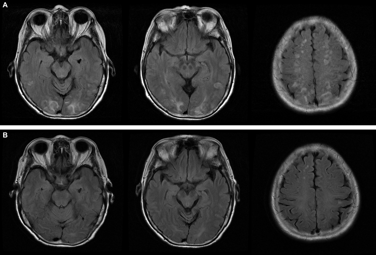
In laboratory test, she showed hyponatremia, which is 124 mmol/L. Cerebrospinal fluid study revealed normal, which was performed to exclude central nervous system infection. Electroencephalography showed 5–6 Hz diffuse background slowing and there was no epileptiform discharges. Brain magnetic resonance imaging (MRI) showed bilaterally diffuse abnormal signal intensities in the parieto-occipital lobe and superior frontal sulcus, which involved predominantly the deep white matter (Fig. 1A). All these changes showed hyperintense on T2-weighted and FLAIR images. There was no enhancement in meninges and brain parenchyme. Her clinical symptoms and brain MRI findings suggested the possibility of PRES.

(A) Axial fluid-attenuated inversion recovery (FLAIR) MR images demonstrate bilateral hyperintense lesions in the parietooccipital and frontal lesions affecting the cortex and subcortical white matter which consistent patterns with vasogenic edema. (B) Axial fluid-attenuated inversion recovery (FLAIR) MR images show that the lesions have much improved after 1 month.
During admission, her blood pressure was normal. She was treated with saline infusion for correction of hyponatremia. After treatment, her consciousness gradually improved. After three days, her sodium level was normal and she did not complain of lethargy or somnolence. She discharged from hospital without neurological deficit. After one month, follow-up brain MRI revealed that abnormal signals involving both parieto-occipital cortex and superior frontal sulcus were much improved (Fig. 1B).
Discussion
Most of the previously described PRES had associated extremely high blood pressure or renal insufficiency.1–3 Other etiologies are known such as pregnancy, immunosuppressant drugs use, nephrotic state, sepsis, autoimmune diseases and CNS infection.5 Various previous studies reported other miscellaneous associations such as hypomagnesemia, hypercalcemia, tumor lysis syndrome, rapid correction of anemia, HIV infection and ESRD.5,6 Numerous aspects regarding the pathogenesis of this entity are yet to be elucidated. Although there are many proposed mechanisms for the patho-physiology of PRES, the most widely accepted is a temporary failure of autoregulatory capabilities of the cerebral vessels, leading to hyperperfusion, a breakdown of the blood brain barrier and consequent vasogenic edema with some component of endothelial dysfunction.5,6
In our case, she had no other etiology such as high blood pressure, but she showed only hyponatremia. Despite the importance of blood vessels function for the homeostasis of the brain, little is known about the influence of hyponatremia on cerebrovascular regulation.7 One previous study demonstrated that the dilatation of the cerebral vessel in response to acetylcholine, which is endothelium and nitric oxide dependent, was severely impaired in hyponatremia.8 Cerebral hypoxia due to vasoconstriction probably makes impairment of brain adaptation due to dysfunction of Na+−K+−ATPase, which is primary importance for the extrusion of ions from the glial cell.9 Also, vasopressin elevated in cases of hyponatremia. Vasopressin leads to decreased cerebral oxygen utilization and facilitates direct movement of water into brain cells independent of the effects of hyponatremia.10 Therefore, these mechanisms in hyponatremia may have an important impact on the brain edema and could explain our case.
To our knowledge, this is the first case of PRES associated with hyponatremia in Korea. We suggest that a rapid change of sodium level may cause disruption of cerebral autoregulation, which result in PRES. While PRES is usually reversible, the early recognition and treatment of this syndrome is important to prevent permanent neurological sequelae. Further investigation is needed to elucidate the association between sodium level and the development of PRES.